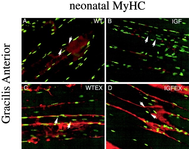
Figure 6.
Neonatal myosin localization on gracilis anterior muscles with Hoechst (green fluorescence) and AChE (brightfield) stains. (A–D) Gracilis anterior with AChE precipitate between arrows. (A) WT, (B) MLC/mIGF-1 transgenic, (C) WT-EX, (D) MLC/mIGF-1 transgenic exercised. Neonatal myosin is upregulated along the length of fibers in WT-EX and IGF-EX muscles.

