Figure 1.
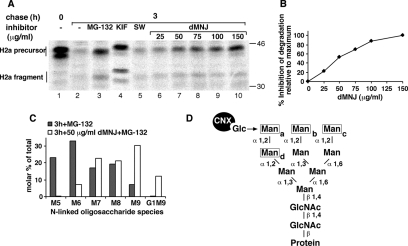
Involvement of a class I α-mannosidase in ERAD of ASGPR H2a. Partial inhibition of mannosidase activity blocks ERAD while still allowing trimming to M8. (A) NIH 3T3 cells stably expressing H2a (2-18 cell line) were pulse labeled with [35S]Cys for 20 min followed by a 3-h chase in complete medium with or without addition of 20 μM MG-132 or of saturating amounts of the mannosidase inhibitors KIF (100 μM) and SW (4 μg/ml) or of the indicated concentrations of dMNJ. (Cells were preincubated for 30 min with the mannosidase inhibitors, which were also present during the labeling period.) Cells were lysed, H2a was immunoprecipitated, and the immunoprecipitates were separated in 10% SDS-PAGE followed by fluorography. Bands corresponding to the H2a precursor and the naturally occurring cleaved fragment are indicated on the left. Molecular mass markers (kilodaltons) are indicated on the right. The bands below the upper H2a precursor and fragment species are underglycosylated molecules (lacking one of the three sugar chains). (B) Percentage of inhibition of degradation of H2a relative to maximum inhibition obtained with 150 μg/ml dMNJ (=100) was calculated from a phosphorimager quantitation of the gel in A. Note that although reaching a plateau, dMNJ only inhibits degradation of H2a by 50% compared with 80% with KIF. (C) The same cells as in A were labeled with 2-[3H]Man for 1 h in glucose-free medium and chased for 3 h in complete medium in the presence of 20 μM MG-132 with or without 50 μg/ml dMNJ. Cells were lysed, H2a was immunoprecipitated, and the immunoprecipitates were treated with endo H. The N-linked oligosaccharides were separated by HPLC, and fractions were counted in a beta counter. Relative molar amounts of each oligosaccharide species were calculated based on mannose content. The percentage of each species relative to the sum of the molar amounts of all species present was then plotted. (D) Structure of M9, highlighting the α1,2-linked mannose (Man) residues (a–d) (boxed) that undergo trimming in the ER and the position (arrow) of the glucose (Glc) residue on the original precursor (after its two outer glucoses were excised) or after its readdition by UGGT. Note that trimming of mannose-c and/or mannose-b does not prevent reglucosylation but loss of mannose-a does.

