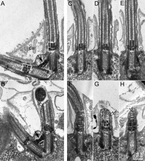
Figure 4.
Ultrastructural analysis of WT and mutant cells reveals TZ defects in uni2 mutants. Thin sections of WT or mutant strains were viewed using TEM. (A and C) WT cells have uniform TZs composed of proximal (p) and distal (d) electron dense cylinders forming an H-shaped region in longitudinal section (white brackets). The flagella project through openings in the cell wall (CW). (B) Mutant uni2-2 cells have the normal arrangement of two basal bodies connected by a distal striated fiber (black arrow) but most basal bodies have a (D) truncated and/or (B) disrupted distal TZ cylinder. (E–H) Mutant uni2-3 cells have (E) elongated distal TZ cylinders or (F–H) extra stacked TZ material. Basal bodies unable to assemble a complete flagellum sometimes show a flagellar stump with some axonemal microtubules (white arrows, B and G). In some cases, the tip of the stump is filled with amorphous material (open arrow, H).

