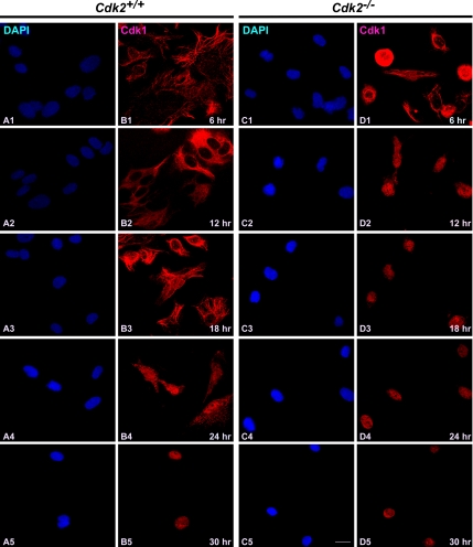
Figure 1.
Cdk1 translocates early to the nucleus in the absence of Cdk2. Immunocytochemical staining of Cdk1 in Cdk2+/+ and Cdk2−/− MEFs at different time points after serum stimulation. Localization of Cdk1 was detected by rabbit anti-Cdk1 antibodies followed by AlexaFluor568 goat anti-rabbit antibodies (red; B1–B5 and D1–D5), and the nuclei were counterstained with DAPI (blue; A1–A5 and C1–C5). The images were captured with a laser confocal microscope (63×). The representative pictures are from one of three independent stainings. Scale bar, 10 μm in every panel.

