Figure 3.
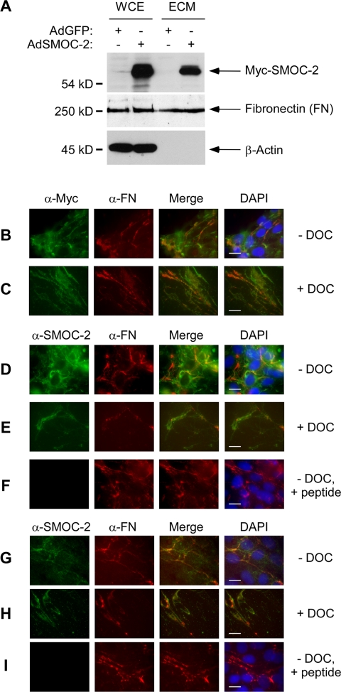
Subcellular distribution of SMOC-2. (A) Whole cell lysates and ECM preparations from AdCon- and Ad-SMOC-2–infected Rat1 cells were resolved by SDS-PAGE, transferred to nitrocellulose and probed with an anti-Myc antibody. The same samples were probed with antibodies against FN and β-actin. AdSMOC-2–infected (B–F) or uninfected (G–I) Rat1 cells were fixed and stained with anti-Myc and anti-fibronectin (B and C) or anti-SMOC-2 and anti-fibronectin (D–I) antibodies. Bound primary antibodies were detected using a FITC-conjugated antibody (for detecting anti-Myc and anti-SMOC-2) and a Cy3-conjugated antibody (for detecting anti-fibronectin). In some experiments, cells were extracted with DOC buffer before fixing and antibody staining (C, E, and H). Some antibody incubations with were carried out in the presence of a competitor peptide corresponding to the epitope recognized by the SMOC-2 antisera (F and I). Bars, 10 μm.

