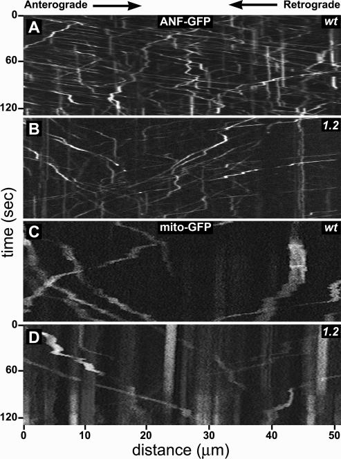
Figure 4.
Live transport behavior of organelles in unc-104 mutant axons. Each panel, extracted from a time-lapse movie of GFP-organelles in motor axons of a larval segmental nerve, shows a kymograph representation of fluorescent organelle positions as a function of time. Anterograde movements have negative slopes, whereas retrograde movements have positive slopes. Stationary organelles appear as vertical streaks. Before each movie, the field of view was photobleached, which reduced signal from stationary organelles, allowing better contrast for organelles that subsequently moved into the bleached area. (A and B) ANF::GFP shows DCV behavior in wild-type (wt) and unc-104O1.2/unc-104P350 (1.2) axons. Note the lower abundance of anterograde DCVs and their slower movements (larger negative slopes) compared with wild type. (C and D) MitoGFP shows mitochondrial behavior. Intact time-lapse movies of organelle transport can been seen in Supplemental Movies S1–S6.

