Figure 1.
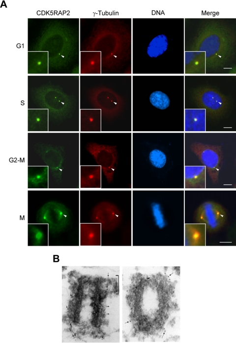
Centrosome localization of CDK5RAP2. (A) HeLa cells were double labeled for CDK5RAP2 and γ-tubulin. Arrow-pointed centrosomes are enlarged in insets. Bars, 10 μm. (B) Electron micrographs representative of isolated HeLa centrosomes after reaction with an antibody to CDK5RAP2 and a gold-labeled secondary antibody. The arrows point to CDK5RAP2 labels, and the bracket indicates a centriolar appendage. Magnification, ×80,000.

