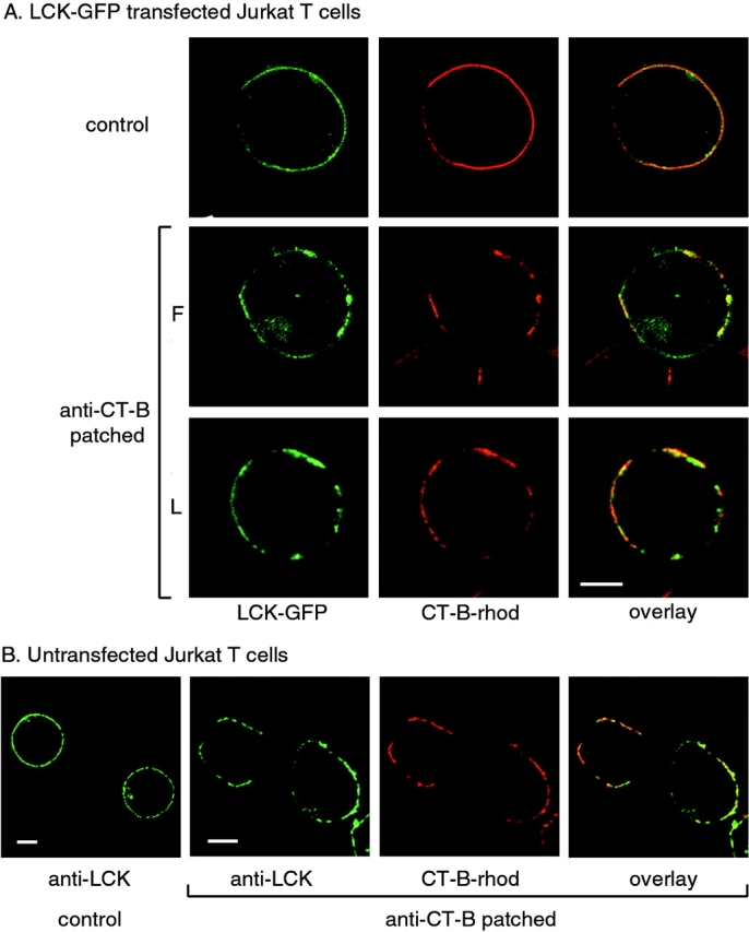
Figure 1.

Subcellular localization of LCK and CT-B–rhodamine in Jurkat T cells. A, Cells transiently transfected with LCK-GFP were incubated with CT-B–rhodamine and left untreated (control) or treated by cross-linking with anti–CT-B antibody, as described in Materials and Methods. Single confocal sections were taken of either fixed (F) or live (L) cells using fluorescence in GFP and rhodamine channels. B, Control and anti–CT-B patched cells were fixed and stained with anti-LCK antibody for endogenous protein and visualized by confocal microcopy as in A. Bars, 5 μm.
