
Figure 5.
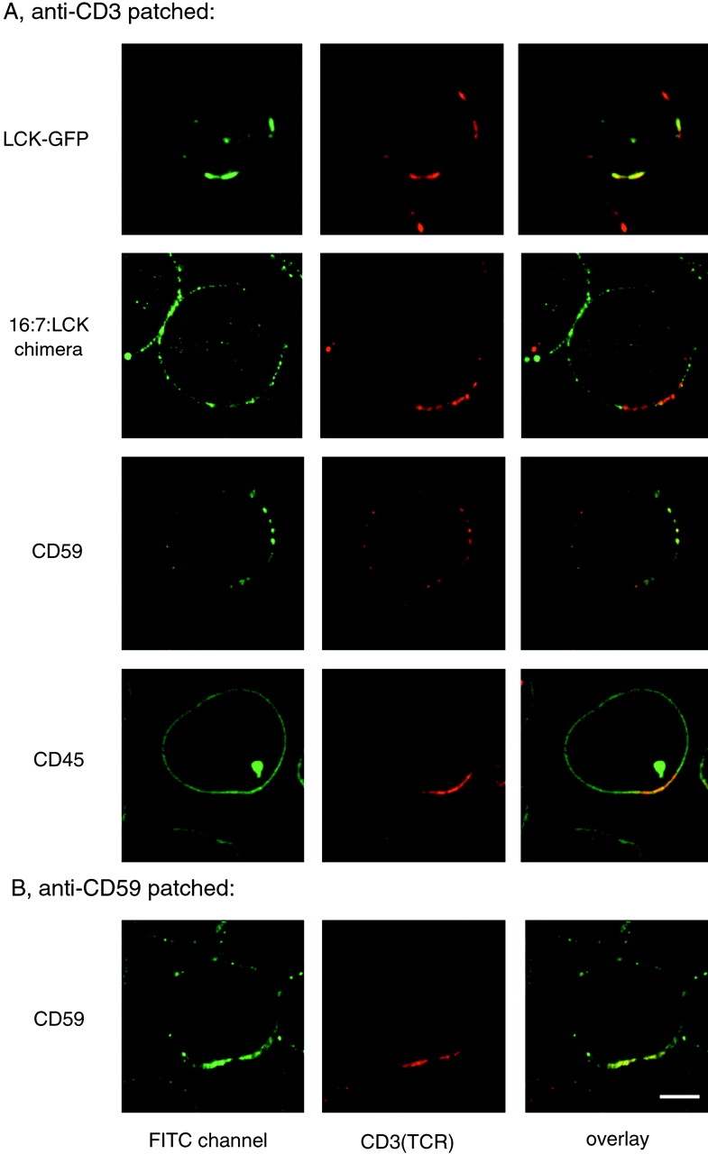
TCR patches resemble lipid raft patches in protein content. A, Cells expressing LCK-GFP (top row), 16:7:LCK (second row), or untransfected Jurkat cells (other rows) were incubated with anti-CD3 mAb followed by anti-Ig Texas red to induce receptor patching. Cells were then fixed and stained as indicated with anti-LCK antiserum followed by FITC-conjugated secondary antibody, or with FITC-conjugated mAbs against CD59 or CD45, to avoid cross-reactivity with anti-CD3 mAb. B, Jurkat cells were incubated with FITC-conjugated anti-CD59 mAb followed by anti-Ig to induce receptor patching. The patched cells were then stained with biotin-conjugated anti-CD3 mAb followed by streptavidin Texas red. Bar, 5 μm.