
Figure 8.
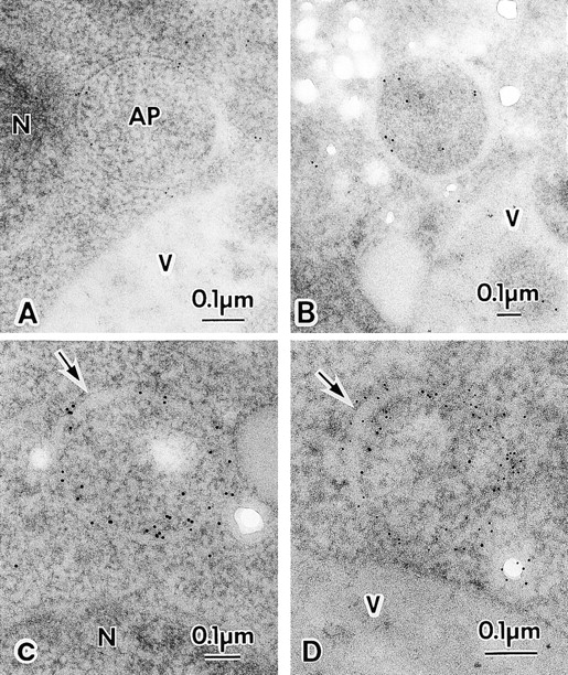
Immuno-EM of autophagosomes. A and B, Mature autophagosome. C and D, Premature autophagosome. Arrows show expanded regions of the intramembrane space of the premature autophagosomes. TK116 cells were incubated in SD(−N) medium for one hour (D), two hours (B and C), or three hours (A). The localization of 3 × HA–Apg8p was detected with anti-HA antibody, followed by the incubation with 5-nm gold- (A and D) or 10-nm gold- (B and C) conjugated goat anti–mouse IgG. AP, autophagosome; N, nucleus; V, vacuole.