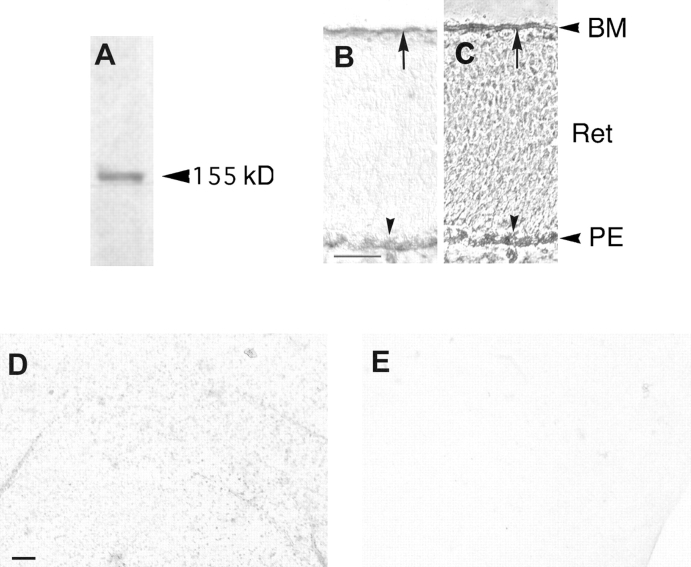
Figure 4.
A shows the α1-AP fusion protein in cos7 cell supernatant, immunodetected with IG2 sera. α1-AP consists of the extracellular domain of CRYPα, which has a molecular weight of ∼90 kD together with the alkaline phosphatase of 65 kD. B and C show binding of the α1-AP fusion protein to a section of the E6 retina, visualized with AP staining. C is the corresponding phase contrast view. Arrows indicate staining in the BM of the retina. The arrowheads indicate the pigment epithelium. D shows binding of the α1-AP fusion protein to a BM from a E7 retina, visualized with AP staining. The stained dots represent glial endfeet. There is no binding of α1-AP to an E7 detergent-washed endfeet-free BM detectable as can be seen in E. Abbreviations: BM, basal membrane; Ret, retinal cell layer; PE, pigmented epithelium. Bars: (B and C) 0.1 mm; (D and E) 0.01 mm.

