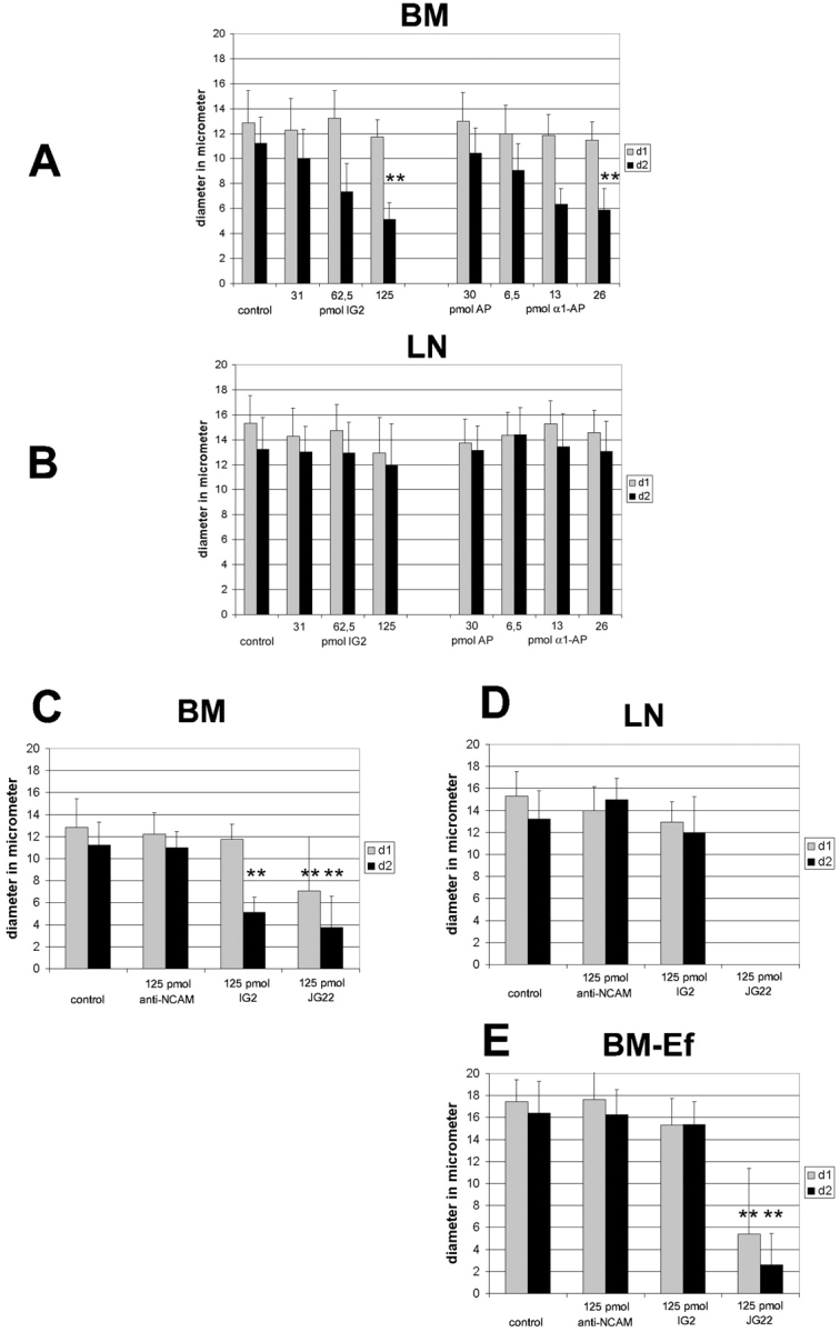
Figure 9.

Quantification of growth cone morphology on different substrates. On BM both IG2 and α1-AP affect growth cone morphology. D2 is significantly reduced in a dose-dependent manner while d1 is not affected at all (A). There was no effect on LN (B). Comparing the influence of both antibody and growth substrates it is remarkable that growth cones on LN and BM-Ef are larger in their overall size compared with ones grown on BM (C–E). Moreover, treatment with IG2 only had a significant effect on d2 of growth cones grown on BM (C), but not on LN (D) and BM-Ef (E). When using 125 pmol JG22 there were not any growth cones detectable on LN (D). Growth cone morphology was less, but still heavily affected on BM-Ef (E) and BM (C) (** indicate statistical significance P < 0.0001). See Table for data.
