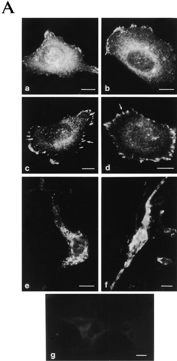
Figure 2.
Characterization of the cellular location of α2 and α2/α1 integrin and formation of actin stress fibers. Stable Saos-2 transfectants were either grown in DME 10% FCS on immunofluorescence glass slides, plated on collagen coated glass slides, and allowed to adhere for 4 h in DME (serum-free) or grown inside a three-dimensional collagen gel for 48 h. (A) After incubation, the cells were fixed and stained for α2 integrin by using mAb followed by FITC-labeled rabbit anti–mouse antibody. Cells were washed, mounted, and examined on a microscope: (a) α2 clone 47 and (b) α2/α1 clone 18 on serum-derived fibronectin and vitronectin; (c) α2 clone 47 and (d) α2/α1 clone 18 on collagen; (e) α2 clone 47 and (f) α2/α1 clone 18 inside collagen gel; and (g) vector clone as a negative control. (B) Cells were stained for actin using fluorescein-conjugated phalloidin (a) α2 clone 47 and (b) α2/α1 clone 18 on collagen.