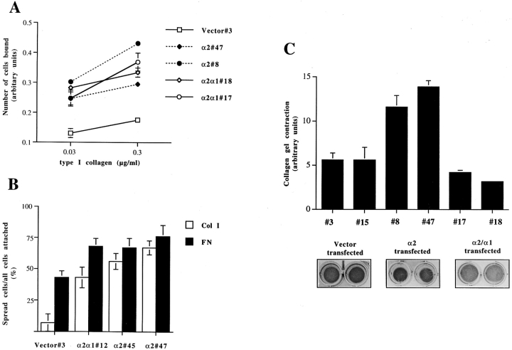
Figure 3.
Cell–collagen interaction of stable transfected human osteosarcoma Saos-2 expressing wild-type α2 integrin, chimeric α2/α1 integrin or empty vector. The data shown are the mean values ± SD of a representative experiment done in triplicate. (A) Cell adhesion to type I collagen was studied. 10,000 cells were suspended in DME (serum-free), then transferred into wells coated with indicated concentrations of type I collagen. After 1 h the nonadherent cells were washed out and the adherent ones were stained with crystal violet. Cell-bound stain was dissolved and measured spectrophotometrically. (B) Cell spreading on type I collagen and fibronectin was studied. 10,000 cells were suspended in DME with 50 μM cycloheximide, and then transferred into wells coated with different matrixes. After 30 min the nonadherent cells were washed out and the adherent ones were fixed. The percentage of spread cells was counted. (C) The ability of various collagen receptor integrins to mediate collagen gel contraction was tested using stable transfected clones of human osteosarcoma Saos-2 cells (mock-, α2-, or α2/α1–expressing clones). 5 × 10⁵ cells were seeded inside a collagen gel, the polymerized gel was detached from the sides of the dish and the cells were cultured for 3 d in DME 10% FCS. The gels were photographed and surface areas of the gels were measured.

