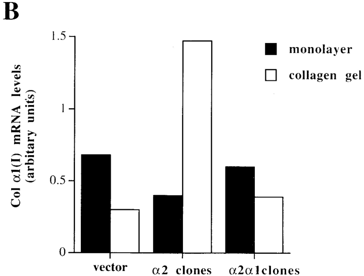
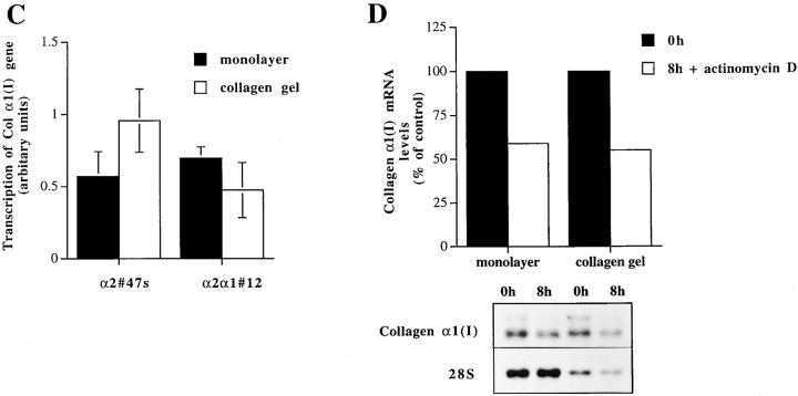
Figure 4.
Effect of matrix on the expression level of collagen α1(I) and collagen α2(I) mRNAs, transcriptional regulation and mRNA stability of collagen α1(I) in Saos-2 cells expressing either wild-type α2 or chimeric α2/α1 integrin. Mock-, α2-, and α2/α1–transfected cells were cultured in a monolayer (M) or inside collagen gel (C) for 48 h. Total RNA was isolated, separated by electrophoresis, and transferred to filters. Specific mRNA levels were analyzed with corresponding cDNA probes. 28S ribosomal RNA was used as a control. (A) Autoradiograms of a representative experiment and (B) quantitative analysis of an identical separately performed experiment was done by an image analyzer. (C) Nuclear run-on assay was performed with nuclei from Saos-2 cells grown either in monolayer or inside three-dimensional collagen gels 48 h before harvest. Nascent 32P-labeled RNA was hybridized to nitrocellulose filter-immobilized cDNA probe. Quantitative analysis was performed with an image analyzer system. The values for the collagen α1(I) gene transcription were corrected to GAPDH transcription in the same sample. The data shown are mean ± range of a representative experiment done in duplicate. (D) mRNA stability assay using actinomycin. Cells were cultured for 24 h before adding actinomycin D (6.4 μg/ml) either inside collagen gel or in monolayer. Total RNA was isolated from cells after 8 h. Autoradiograms from Northern blot analysis using collagen α1(I) cDNA and 28S cDNA as control are shown. Quantitative analysis of the experiment was done with an image analyzer system.


