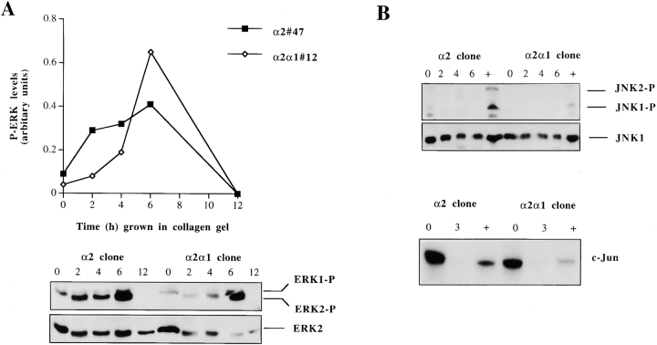
Figure 8.
Regulation of ERK and JNK MAPKs in response to collagen gel. Cells from two separate α2 or α2/α1 single cell clones (45, 47 and 2, 12) were pooled together and either lysed immediately (0 h sample) or seeded in collagen gel and incubated for different periods of time, as indicated. (A) The levels of activated ERK1 and 2 (ERK1-P and ERK2-P) were determined by Western blot analysis using a phosphospecific antibody for ERK1 and 2 and an antibody recognizing all forms of ERK2 was used as a control. The levels of activated ERK1 and 2 were quantitated using an image analyzer system and are shown relative to the levels of total ERK2 present in each sample. (B) The levels of activated JNK1 and 2 were determined by Western blot analysis using a phosphospecific antibody for JNK1 and 2, and an antibody recognizing all forms of JNK1 was used as a control. The positive control treatment (+) was 10 min anisomycin (10 μg/ml). For the kinase assay, equal numbers of stable transfected Saos-2 cells (α2 or α2/α1) were either lysed immediately after trypsinization, treated with collagen for 3 h or anisomycin (10 μg/ml) for 10 min, and the JNK kinase activity was assayed.

