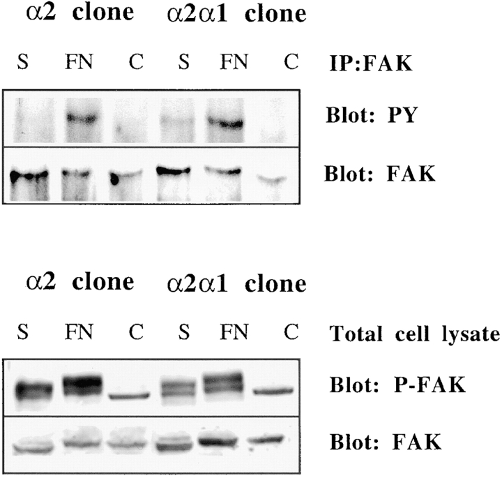
Figure 9.
Three-dimensional collagenous matrix fails to activate FAK in Saos-2 cells. Cells from two separate α2 or α2/α1 single cell clones (45, 47 and 2, 12) were pooled together and either lysed immediately (0 h sample), seeded in collagen gel, or allowed to adhere to fibronectin for 1 h. FAK was immunoprecipitated and immunoplotted with an antiphosphotyrosine antibody (4G10). Alternatively, cell lysates were fractionated on SDS-PAGE and immunoblotted with phosphospecific FAK antibody. The blots were reprobed with anti–FAK antibody to show the levels of FAK protein. Note that in the lower panel the anti-phospho FAK antibody recognizes two bands, but it is the upper one that comigrates with the band recognized with the anti–FAK antibody. The origin of the lower band is unknown.

