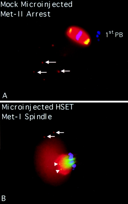
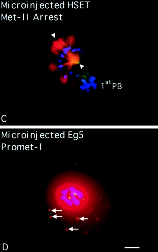
Figure 3.
HSET is essential for microtubule organization in metaphase II spindles in mouse oocytes. Mouse oocytes at the germinal vesicle stage were either mock injected (A), injected with antibodies specific for HSET (Β and C), or injected with antibodies specific for Eg5 (D). Injected oocytes were then matured in vitro until metaphase I (7 h; B) or until metaphase II arrest (16 h; A, C, and D). Oocytes were processed for indirect immunofluorescence using antibodies specific for tubulin (red), DNA (blue), and either Eg5 (A) or the injected antibody (B–D; green). Arrows indicate cytoplasmic asters; arrowheads indicate foci of HSET antigen; and the first polar bodies are marked when discernible (1stPB). E, Percentage of microinjected oocytes that mature to metaphase II arrest after 16 h of maturation in vitro postinjection. Oocytes were either mock injected, injected with antibodies specific for Eg5 antibody, or injected with antibodies specific for HSET, as indicated. Bar, 10 μm.