
Figure 6.
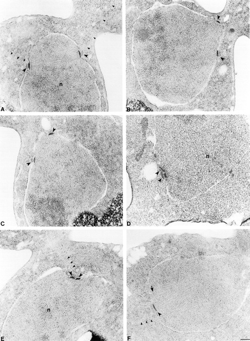
Electron microscopic analysis of kar3-64 kip3Δ cells. Wild-type (A) and kar3Δ kip3Δ (pkar3-64) cells (B–F) were arrested with hydroxyurea at 26°C, shifted to 35°C for 3 h, fixed, and sectioned for electron microscopy. A shows a normal preanaphase bipolar spindle. All the mutant spindles (B–F) appeared defective. In B and C, spindles have lost bipolarity. Although the poles are still slightly separated, they are not parallel. In D, E, and F spindles have collapsed with poles in a side by side orientation. All SPBs are marked with large arrowheads. Arrow in F marks a pole whose edge was just caught in this section. Small arrowheads mark cytoplasmic microtubules. n, nucleus. Bar, 0.2 μm. Strains used are listed in legend of Fig. 1.