
Figure 2.
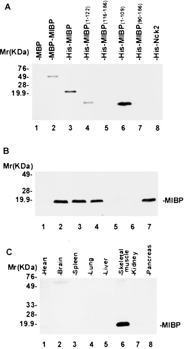
MIBP is predominantly expressed in skeletal muscle. (A) Immunoblot with mAb 5B4.7. Each lane was loaded 10 ng of recombinant proteins. Lane 1, MBP; lane 2, MBP–MIBP; lane 3, His–MIBP; lanes 4–7, His fusion proteins containing partial MIBP sequences as indicated in the figure; and lane 8, His fusion protein containing an irrelevant protein Nck2. (B) Immunoblot showing mAb 5B4.7 specifically recognizes mammalian MIBP. Equal amounts of C2C12 cell extracts (5 μg/lane) were probed with 3.3 nM irrelevant control mouse IgG (lane 1), 3.3 nM mAb 5B4.7 (lane 2), 3.3 nM mAb 5B4.7 preincubated with 1.65 nM (lane 3), 3.3 nM (lane 4), 16.5 nM (lane 5), or 33 nM (lane 6) MBP–MIBP, or 3.3 nM mAb 5B4.7 preincubated 33 nM MBP (lane 7). Note that binding of mAb 5B4.7 to mammalian MIBP was blocked by an excess amount of MBP–MIBP (lanes 5 and 6) but not of MBP (lane 7). (C) MIBP is predominantly expressed in skeletal muscle. Equal amounts (15 μg/lane) of human fetal tissues were analyzed by immunoblotting with mAb 5B4.7.