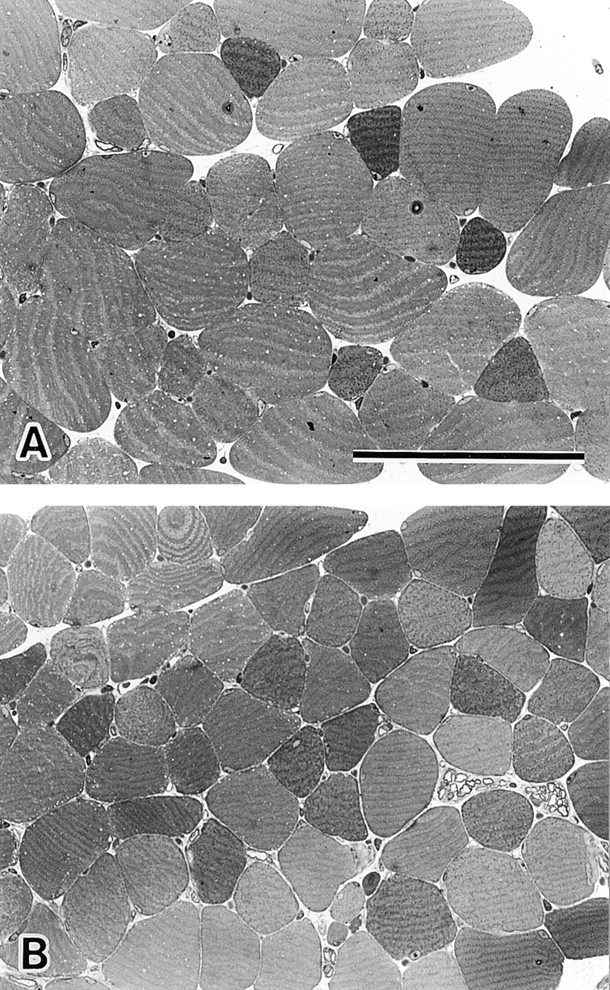
Figure 3.

Histological analysis of MG29-deficient EDL muscle. Toluidine blue–stained cross sections of EDL muscles from hind limb of 8-wk-old wild-type (A) and MG29-deficient mice (B) are shown. The cross-sectional areas of mutant muscle cells are significantly reduced compared with those of the control muscle cells. However, no significant differences between the genotypes are observed in the number of muscle cells and myofibril density within the cells. Bar, 100 μm.
