
Figure 4.
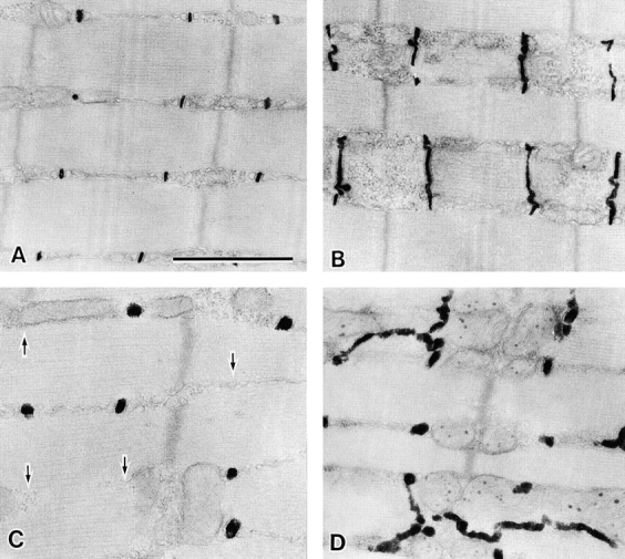
Abnormal T tubular system in MG29-deficient skeletal muscle. Electron micrographs of the longitudinal sections of T tubule–stained hind limb muscles from 8-wk-old wild-type (A and B) and MG29-deficient (C and D) mice. The stained T tubules are swollen and run in irregular directions in the mutant muscle cells. Arrows in C indicate the missing T tubules at the A-I junctions. Bar, 1 μm.