
Figure 2.
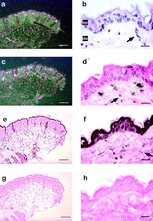
Cell type-specific expression of MMP-13, MMP-3, and GR in skin of TPA-treated mice. In situ hybridization for MMP-13 (a and b) and MMP-3 (c and d) on transverse sections of the skin from female C57BL/6 mice treated for 6 h with TPA. Dark-field (a) and bright-field (b) illumination of a section hybridized with a MMP-13 specific riboprobe. The arrow indicates nuclei from fibroblasts. Dark-field illumination (c) and bright-field (d) illumination of a skin section hybridized with a MMP-3 specific riboprobe. e–h, Immunohistochemical analysis of GR expression using anti-mouse GR antibody (e and g) or secondary anti-rabbit IgG antibody alone (g and h). The arrow indicates nuclei from fibroblasts. ep, Epidermis; de, dermis. Bars: (a, c, e, and g) 100 μm; (b, d, f, and h) 20 μm.