
Figure 5.
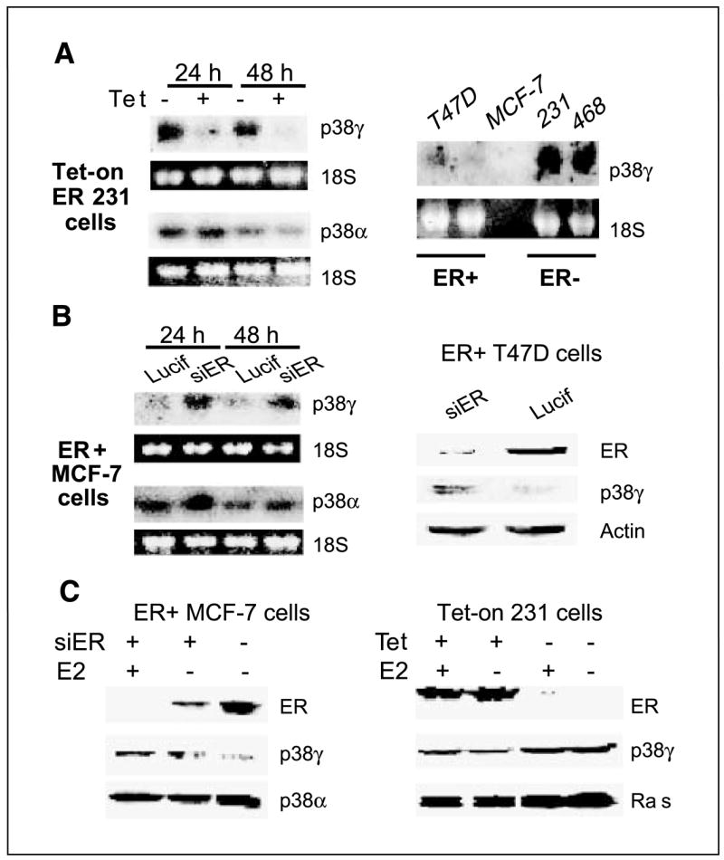
ER negatively regulates p38γ expression in a panel of human breast cancer cell lines. A, ER+ phenotype correlates with lower levels of p38γ RNA expression. Total RNA was prepared, separated, and transferred to a nitrocellulose membrane that was hybridized with a specific radioactive-labeled probe for p38γ expression. B, ER depletion increases p38γ expression. ER+ cells were infected with pSR-Lucif (Lucif) or pSR-siER (siER) for 24 hours and examined for p38γ RNA (left) or protein (right) expression. C, ER negatively regulates p38γ expression by ligand-independent mechanisms. Cells in steroid-depleted medium were incubated for 24 hours (±10 nmol/L E2) after the pSR infection (left) or in the presence or absence of Tet (right) and analyzed by Western analysis for protein expression.