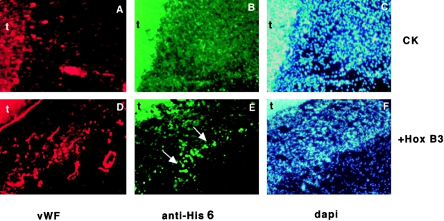
Figure 7.
Increased capillary density correlates retrovirally expressed Hox B3. Immunofluorescence staining of serial 7-μM sections of CAMs harvested 72 h after application of fibrosarcoma cells producing empty retrovirus (CK) or retrovirus expressing Hox B3 with a His-6 epitope tag fused to the COOH terminus (+Hox B3). Staining with an antibody against von Willebrand factor (A and D) shows a relative increase in endothelial cell density in membranes exposed to Hox B3. Staining of serial sections with an antibody against the 6×His epitope tag fused to Hox B3 (B and D) reveals positive staining (arrows) in areas associated with increased vascular density. Areas showing strong autofluorescence within the tumor cores are indicated by the letter t. Corresponding DAPI nuclear staining is shown for both tissues (C and E).

