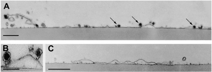
Figure 6.
Cross-sections of membrane patches analyzed by electron microscopy. The fragments were incubated either in the presence of 2 mM EGTA (A) or in the presence of 100 μM Ca2+ (C) for 3 min before fixation. Several secretory vesicles (arrowheads) are attached to the cytoplasmic face of the plasma membrane in the EGTA-treated patch. (B) Two secretory vesicles at higher magnification. Bars: (A and C) 0.5 μm; (B) 0.25 μm.

