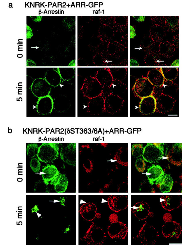
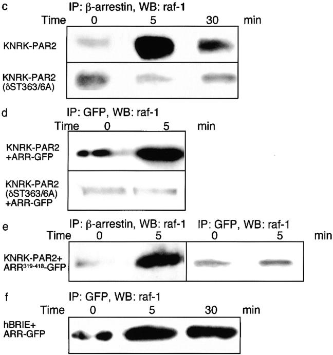
Figure 7.

Trypsin induced association of β-arrestin and raf-1. (a and b) Localization of β-arrestin and raf-1 by immunofluorescence and confocal microscopy. KNRK-PAR2+ARR-GFP cells (a) or KNRK-PAR2(δST363/6A)+ ARR-GFP cells (b) were incubated with 50 nM trypsin for 0 or 5 min at 37°C. β-Arrestin was localized using GFP and raf-1 was localized by immunofluorescence. The same cells are shown in each row and the images in the right panel are formed by superimposition of images from the other two panels in the same row. Representative of two experiments. In KNRK-PAR2+ARR-GFP cells, note the redistribution of β-arrestin and raf-1 from the cytosol at 0 min (arrows) to the plasma membrane at 5 min (arrowheads), where they colocalize. In KNRK-PAR2 (δST363/6A)+ARR-GFP cells, note that β-arrestins remain in the cytosol (arrows) and raf-1 redistributes to the plasma membrane at 5 min (arrowheads). (c–f) Coimmunoprecipitation of raf-1 and β-arrestin. Cells were incubated with 50 nM trypsin for 0–30 min at 37°C, lysed, immunoprecipitated (IP) using antibodies to GFP or β-arrestin-1/2, and analyzed by Western blotting (WB) with a raf-1 antibody. (c) In KNRK-PAR2 cells, but not in KNRK-PAR2(δST363/6A) cells, β-arrestin and raf-1 coprecipitated. (d) Similarly, in KNRK-PAR2+ARR-GFP cells but not KNRK-PAR2(δST363/6A)+ARR-GFP cells ARR-GFP and raf-1 coprecipitated with antibodies to GFP. (e) In KNRK-PAR2+ARR319-418-GFP cells, endogenous β-arrestin and raf-1 coprecipitated, but ARR319-418 and raf-1 did not coprecipitate. (f) In hBRIE+ARR-GFP cells, ARR-GFP and raf-1 coprecipitated. Bars, 10 μm.

