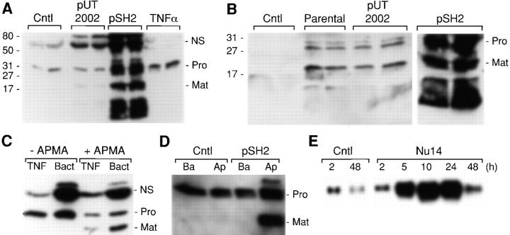
Figure 1.
FimH-dependent induction and activation of matrilysin in epithelial cells. (A) WiDr human colon carcinoma cells were infected for 90 min with E. coli strains ORN103/pUT2002 (type 1+/fimH −) and ORN103/pSH2 (type 1+/fimH +) at a ratio of 300:1 bacteria/epithelial cells. Cultures were washed extensively and re-fed with medium containing 50 μg/ml gentamicin. Some cultures were treated for 48 h with 50 ng/ml of human recombinant TNFα. Conditioned media were collected 48 h after infection and analyzed by Western blotting for matrilysin protein. the 28-kD proform of matrilysin (Pro), the 19-kD activated form of matrilysin (Mat), and autolytic fragments (broad migrating faster below activated matrilysin) were seen in pSH2-infected cells. A nonspecific (NS) band is seen at ∼70 kD. Numbers on the left side of the autoradiogram indicate the migration of molecular mass standards. Samples from two separate cultures were analyzed for each group. (B) J82 human bladder carcinoma cells were infected for 90 min with the parental E. coli strain ORN103 and the recombinant strains ORN103/pUT2002 and ORN103/pSH2. Conditioned media were collected 48 h later, and the expression of matrilysin was analyzed by Western blotting. (C) Aliquots of conditioned media from TNFα-stimulated (TNF) or ORN103/pSH2-infected (Bact) colon epithelial cells were incubated at 37°C for 1 h in the presence or absence of 1 mM APMA, and the products were analyzed by Western blotting. (D) WiDr colon cells were grown on Transwell inserts, and some cultures were infected with ORN103/pSH2. Conditioned media was collected after 48 h from the apical and basal compartments and analyzed by Western blotting. (E) HT29 colon cells were infected for 90 min with E. coli NU14 (300 bacteria per epithelial cell). Total RNA was isolated at the indicated times after the infection. Matrilysin mRNA was detected by Northern hybridization.

