
Figure 2.
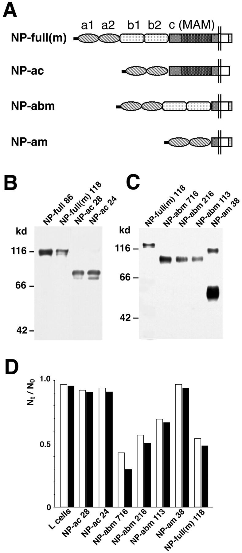
Cell adhesion activity of transfectants which express mutant neuropilin-1 proteins (Part 2). (A) A schematic representation of mutant neuropilin-1s; NP-full(m), full-length neuropilin-1 containing myc tag at the COOH-terminal end; NP-ac, neuropilin-1 lacking the b1 and b2 domains; NP-abm, neuropilin-1 lacking the c domain (myc-tagged); NP-am, neuropilin-1 lacking the b1, b2, and c domains (myc-tagged). (B and C) Immunoblot of the transfectants with the antibody raised against the b1-c domains (B) and anti-myc antibody (C). The number followed by the name of the construct represents the clone number. A band at the 100-kD position in the lane of NP-am appears to be dimerized proteins. (D) Quantification of cell aggregation activity of the mutant neuropilin-1s. White and black bars represent N30/N0 and N60/N0, respectively.