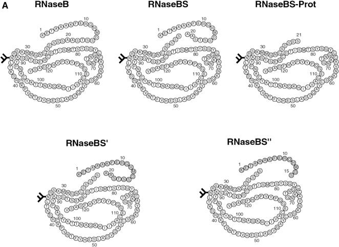
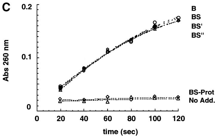
Figure 1.
RNaseB conformers. (A) Schematic representation of some of the RNaseB conformers generated including RNaseB, RNaseBS, RNaseBS-Prot, RNase-B5′ and RNase-B5′′. Shaded parts depict peptides added to regenerate RNase-B5′, and RNase-B5′′. (B) RNase conformers analyzed by SDS-PAGE were as follows: (A) native intact RNaseB, (B) reduced and alkylated RNaseB, (C) RNaseBS, (D) RNaseBS-Prot, (E) RNaseBS′, (F) RNaseBS′′, (G) α-mannosidase–digested RNaseBS-Prot, (H) EndoH-digested RNaseBS-Prot, (I) reduced and alkylated RNaseA, (J) RNaseS, and (K) RNaseS-Prot. (C) Ribonuclease activity of the indicated RNaseB conformers. “No Add.” indicates reactions where no enzyme was added.


