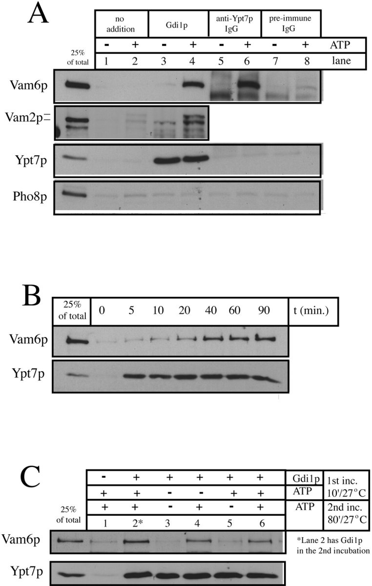
Figure 6.

Release of Vam2/6p from the vacuole. (A) Vam2/6p release. BJ3505 HA-Vam6p vacuoles (see Materials and Methods) were incubated under standard fusion conditions with or without ATP at 27°C. Reactions were supplemented with control buffer, Gdi1p (64 μg/ml), IgG against Ypt7p, or preimmune IgG. After 90 min, the reactions were diluted fivefold with PS buffer and the vacuoles sedimented (5 min, 14,000 g, 4°C). The supernatants were then TCA-precipitated and analyzed by SDS-PAGE and immunoblotting with anti-HA mAb 12CA5 or antisera against Vam2p, Ypt7p, and Pho8p. Lanes 5–8 of the Vam2p panel were omitted because the immunoblot of Vam2p is obscured by the rabbit IgG added to the reactions. (B) Vam6p is released from the vacuole after the release of Ypt7p. BJ3505 vacuoles were incubated under standard fusion conditions with ATP at 27°C in the presence of Gdi1p (64 μg/ml). At the indicated times, reactions were placed on ice. After sedimenting as above, supernatants were analyzed by SDS-PAGE and immunoblotting with antibodies to Vam6p and Ypt7p. (C) The ATP requirement for Vam6p release is unrelated to the release of Ypt7p. BJ3505 vacuoles were incubated at 27°C for 10 min with ATP and/or Gdi1p (64 μg/ml) as indicated. The reactions were then diluted 10-fold with PS buffer and the vacuoles reisolated to remove ATP and Gdi1p. Vacuoles were then resuspended in reaction buffer with or without ATP as indicated and incubated at 27°C for an additional 80 min. Reactions were then analyzed for release of Vam6p and Ypt7p as above.
