
Figure 1.
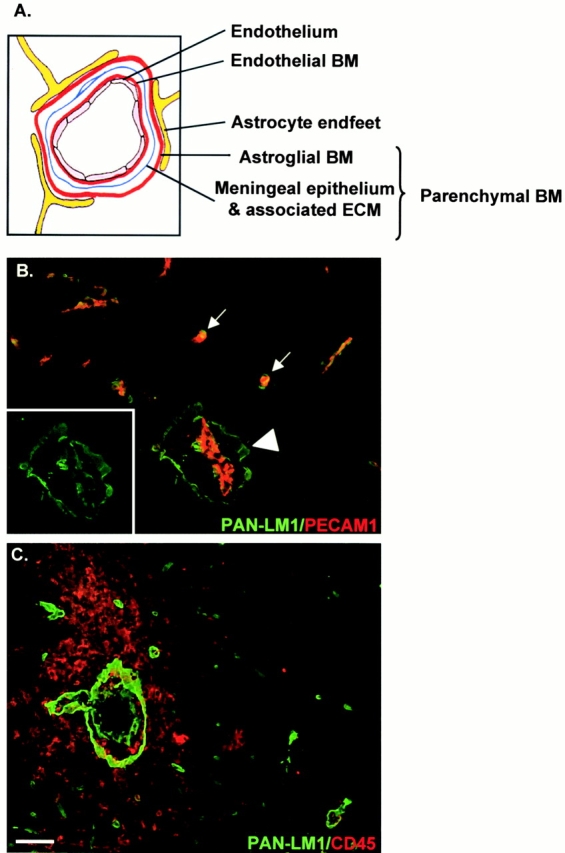
Cell layers and basement membranes which occur in association with blood vessels in the CNS. (A) Schematic representation of blood vessels showing the inner endothelial cell layer and its basement membrane (BM), bordered by epithelial meningeal cells and associated extracellular matrix and outer astroglial basement membrane and astrocyte endfeet. Collectively, the meningeal component and astroglial basement membrane are termed the parenchymal basement membrane. (B) Double staining of EAE brains (clinical scores +1 to +2) for PECAM-1 (red) and pan–laminin 1 antibody (PAN-LM1, green) which recognizes laminin α1, β1, and γ1 chains and thereby many laminin isoforms. Inset shows pan–laminin 1 staining alone. At sites of mononuclear infiltration the internal endothelial cell layer, bordered by its basement membrane, is distinct from the outer parenchymal basement membrane (arrowhead), whereas where no infiltration has occurred these two basement membranes are indistinguishable (arrows). (C) Double staining of EAE brains (clinical scores +3 to +4) with the leukocyte marker, CD45 and pan–laminin 1 antibody, demonstrating infiltrating mononuclear cells accumulating in the perivascular space and penetrating the outer parenchymal basement membrane. Bar, 50 μm.