
Figure 4.
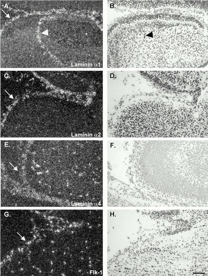
In situ hybridization of consecutive sections of E16 embryonic mouse brains with probes specific for laminin α1 (A and B), α2, (C and D), α4 (E and F), and an endothelial cell–specific probe, flk-1 (G and H). Data shown are for similar areas, encompassing meninges (arrows) and a portion of the cortex of the cerebellum. Similar results were observed for E18 embryos and, in the case of laminin α4, also for newborn and adult tissues. Laminin α1 mRNA was expressed by the leptomeningeal cells of the pia mater that encase the brain and are infolded from the brain surface (arrowheads in A and B); laminin α2 mRNA was restricted to the meninges (C and D), whereas laminin α4 mRNA was detected in endothelial cells within and outside the brain (E and F) in a similar pattern to that observed with the endothelial cell–specific probe, flk-1 (G and H). Bar, 25 μm.