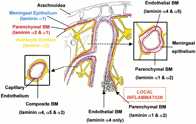
Figure 8.
Illustration of cell layers, basement membranes (BM), and their laminin composition of CNS blood vessels with and without an inflammatory cuff. Larger blood vessels consist of an inner endothelial cell layer with a basement membrane (containing laminins α4 and α5), bordered by the meningeal epithelium and its basement membrane (containing laminin α1) and an outer astroglial basement membrane (containing laminin α2) and astrocyte endfeet. The meningeal and astroglial basement membranes are collectively termed the parenchymal basement membrane as they delineate the border to the brain parenchyma. Only at sites of local inflammation are the endothelial and parenchymal basement membranes distinguishable and define the inner and outer limits of the perivascular space where leukocytes accumulate before infiltrating the brain parenchyma. Examination of such sites demonstrates that mononuclear infiltration occurs across endothelial basement membranes containing only the laminin α4 and bordered by a parenchymal basement membrane containing laminin α1 and α2. The basement membrane of microvessels where no epithelial meningeal contribution occurs appear to have a composite basement membrane containing the endothelial cell laminins, laminin α4 and α5, and laminin α2 produced by the astrocytes and deposited at their endfeet.

