
Figure 1.
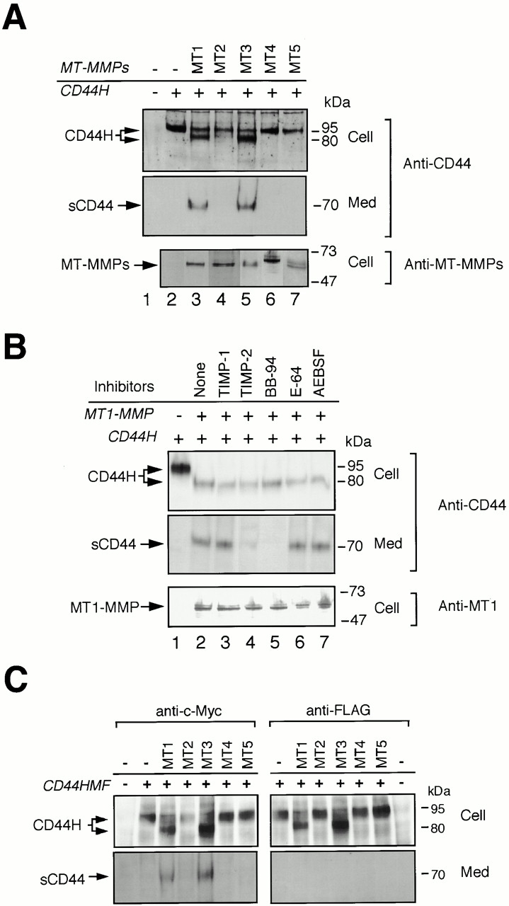
Shedding of CD44H by MT-MMPs. (A) CD44H was coexpressed with each of the MT-MMPs, as indicated by transient transfection of the expression plasmids into ZR-75-1 cells, and incubated in the serum-free media. After 48 h, cell lysates and medium fractions were collected and subjected to Western Blot analyses using monoclonal anti-CD44 and specific antibodies against each MT-MMP. (B) ZR-75-1 cells were transiently transfected with the expression plasmids for CD44H and MT1-MMP and cultured in serum-free media in the presence or absence of various proteinase inhibitors as indicated. After 48 h, cell lysates and medium fractions were collected and subjected to Western blot analyses. (C) CD44H with NH2-terminal c-Myc tag and COOH-terminal FLAG tag was coexpressed with each of the MT-MMPs, as indicated by transient transfection of the expression plasmids into ZR-75-1 cells, and analyzed the same as in A. The antibody against FLAG and c-Myc were used to determine the integrity of the peptide core of CD44H for the Western blot as indicated.