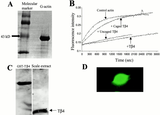
Figure 1.
(A) SDS-PAGE of purified fish actin (right lane) after Coomasie blue staining. The presence of only one major band at the 43-kD marker position (left lane) confirms the purity of extracted actin. (B) Spectrofluorophotometric data from in vitro acrylodan actin polymerization assay under different conditions as indicated, demonstrating the loss of actin-sequestering capability of Tβ4 as a result of caging with NVOC (molar ratio of caged Tβ4/actin was equal to 4:1). Uncaging restored the biochemical activity of caged Tβ4, comparable to the efficiency of pure Tβ4. (C) Western blot of keratocyte scale extracts with anti-Tβ4 antibody (right lane). GST-Tβ4 served as a positive control for the antibody binding (left lane). (D) Immunostaining of Tβ4 in keratocytes demonstrating its diffuse intracellular distribution.

