
Figure 5.
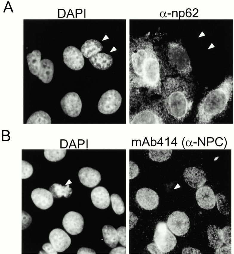
Nuclear pores in apoptotic cells are not detectable by antibodies to nuclear pore proteins. MCF-7 were treated with 50 μM cisplatin for 10 h, fixed, and stained with DAPI to visualize chromatin and with antibodies to nuclear pore protein np62 (A) or monoclonal antibody 414, which reacts with multiple nuclear pore proteins (B). White arrows indicate cells that have lost nuclear pore staining and have condensed chromatin.