Figure 2.
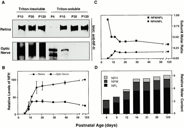
Regional differences in NFH protein and neurofilament subunit stoichiometry during postnatal development. (A) Western blot analysis of Triton-soluble fractions of the retina and optic nerve with the anti-NFH antibody, SMI33. (B) Relative levels of total NFH protein in Triton-insoluble fractions of optic nerve (•) and intraretinal optic axons (▪) were quantified by densitometry after immunostaining with SMI33. Levels of NFH immunoreactivity are expressed relative to that in the adult optic nerve given a value of 100%. Error bars indicate SEM (n = 4 at each age). (C and D) The ratios of neurofilament subunits in Triton-insoluble cytoskeletons of the optic nerve during postnatal development, determined by quantitative Western blot analysis (see Materials and Methods). (C) Molar ratios of NFH to NFL and NFM to NFL at different postnatal ages. (D) The relative subunit molar ratio at different postnatal ages expressed relative to the values at P120. Relative levels of NFH, NFM, and NFL immunoreactivity were calculated from optic nerves at P120 and adjusted to the neurofilament subunit molar ratio determined after metabolic radiolabeling of optic axon proteins in vivo (NFH/NFM/NFL = 1:2:6) as described previously (Nixon and Lewis 1986).

