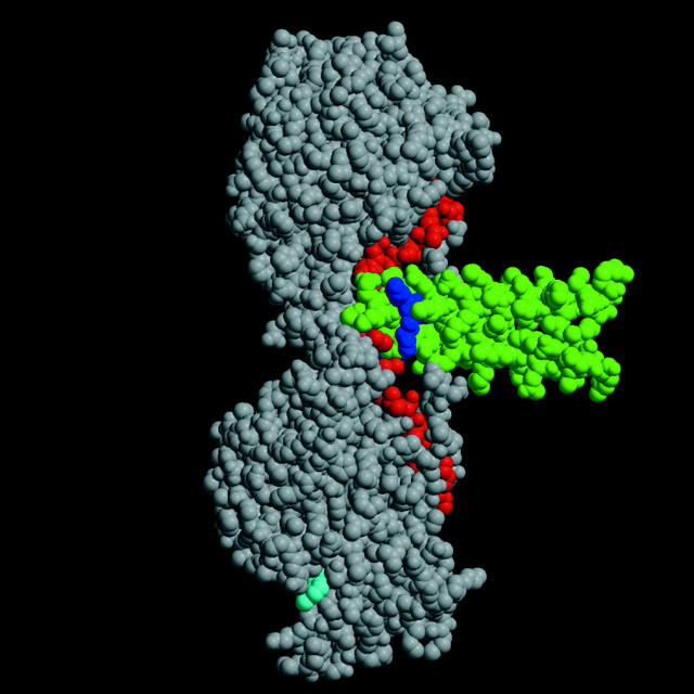
Figure 1.
Kinesin dimer structure. The structure of the kinesin dimer (Kozielski et al. 1997) with the catalytic core shown in gray, the neck linker in red, and the neck coiled-coil in green. The bound ADP is shown in cyan. The two terminal lysines of heptad one are shown in blue.

