Figure 6.
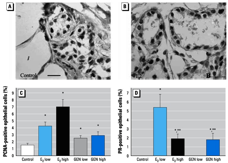
Results of immunostaining for PCNA (A,C) and PR (B,D) in mammary gland. (A) Photomicrograph showing PCNA immunostaining in control tissue. (B) Photomicrograph showing PR immunostaining in E2 high mammary gland. Percentage of PCNA-positive (C) and PR-expressing (D) epithelial cells in mammary terminal structures. Immunostained PCNA cells were regularly seen in all animals (C). PR-expressing cells were seen in both in E2 groups and in the GEN high group (D); note the reduced number of PR-expressing cells in E2 high versus E2 low mammary glands. Bars = 20 μm.
*p < 0.05 compared with control; **p < 0.05 compared with E2 low.

