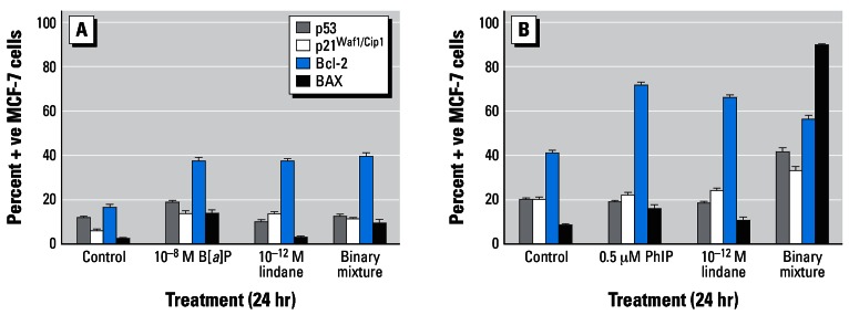
Figure 7.
Immunocytochemical analysis of MCF-7 breast cells treated with (A) B[a]P and/or lindane or (B) PhIP and/or lindane. Cells were treated as indicated for 24 hr on coverslips, after which they were analyzed for protein expression as described in “Materials and Methods.” The percentages of cells staining positive were determined after five separate counts of 100 cells and are presented as the mean ± SD.

