
Figure 5.
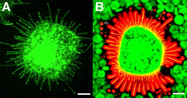
Hyaluronan coat on MCF-7 cells expressing GFP-HAS3. The microvilli (with green GFP-Has3), shown alone in (A), are actually covered by a 0.5–2 μm layer of hyaluronan (B), visualized by a probe made of aggrecan G1 domain and link protein tagged with Alexa Fluor 594® (red). Note that the hyaluronan coat visualized by red blood cells (green) corresponds to the space occupied by the microvilli (yellow) and their hyaluronan cover. The image represents a single confocal optical section; many of the microvilli are shown in cross section (arrow). Bar equals 10 μm.