
Fig. 2. Scanning electron microscopic and histological analyses of palates from wildtype, Tgfb3-Cre homozygote, and Tgfb1 knockin homozygote embryos.
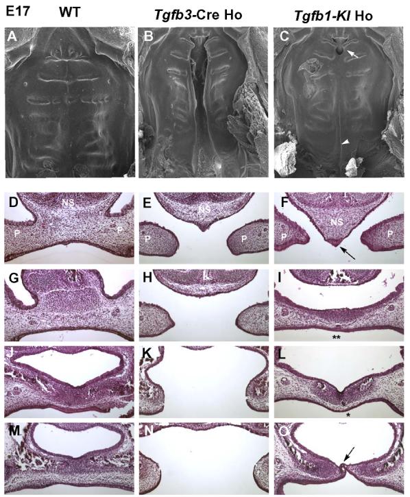
(A-C) Scanning electron microscopic images of palate from wildtype (WT), Tgfb3-Cre homozygote (Tgfb3-Cre Ho), and Tgfb1 knockin homozygote (Tgfb1KI Ho) mice at E17. A wildtype specimen displays a fully fused palate. Tgfb3-Cre homozygote mouse exhibits a complete bilatateral clefting of the secondary palate, whereas the Tgfb1-KI homozygote mouse only has a cleft on the junction of primary and secondary palate (arrow) and a submucous cleft in the posterior region (arrowhead). (D-O) Samples from E17 embryos were sectioned serially in the frontal orientation and stained by hematoxylin-eosin. Sections at four different levels are shown for wildtype (D, G, J, M), Tgfb3-Cre Ho (E, H, K, N), and Tgfb1-KI Ho (F, I, L, O) embryos. In the Tgfb1-KI homozygote sample, the nasal septum fails to fuse with palatal shelves in the anterior region of the palate (F, arrow); palatal shelves fused in the middle region of the palate with the epithelial islands (I, L; single and double stars), indicating an ongoing process of palatal fusion; a submucous cleft is present in the posterior region of the palate (O, dotted arrow). In the wildtype sample, the palatal shelves demonstrate a complete fusion. In the Tgfb3 knockout sample, the palatal shelves display a failure to fuse along the anterior-posterior axis. NS: nasal septum; P: palatal shelf.