
Fig. 4. Comparison of the phospho-Smad2/3 state in the midline palatal epithelium.
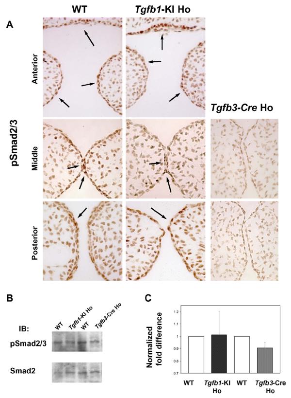
A, Paraffin sections from wild-type (left panel), Tgfb3-Cre homozygote (Tgfb3-Cre Ho, right panel), and Tgfb1-KI homozygote (Tgfb1-KI Ho, mid panel) were immunostained with a phospho-specific antibody against phosphorylated Smad2 and Smad3 (anterior to posterior from top to bottom). In wild-type and Tgfb1-KI Ho samples, cells in the periderm, palatal epithelium and in the nasal septum (epithelium) display a strong positive signal (arrows left and mid panels), whereas in the Tgfb3-Cre sample (right panel) the pSmad2/3 staining is undetectable (n=3).
B, Tissues harvested from tips of prefusion palatal shelves were analyzed for Smad2/3 phosphorylation using western blotting with anti-phopho-Smad2/3 antibodies. C, The histogram represents the relative quantitation of the scanned images. The wild-type (control) phospho-Smad2/3 to Smad2/3 ratio was arbitrarily set at 1 (n=3).