
Fig. 6. Histological analysis and TUNEL assays on palatal shelves from control, Tgfb3-Cre homozygote, and Tgfb1-KI homozygote mice.
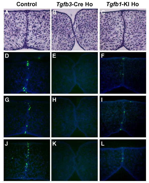
Embryos of each genotype indicated were harvested at E14 and sectioned serially (frontal orientation). (A-C) Histological analysis of frontal sections at comparable levels, staining with hematoxylin & eosin. Palatal shelves of Tgfb3-Cre homozygote made contact but failed to adhere and form epithelial triangles (B). In contrast, palatal shelves of the Tgfb1-KI homozygote (C) were adherent and formed epithelial triangles, which were similar to those from the control sample (A). (D-L) TUNEL assays on frontal sections at three different levels from each genotype (n=3). The Tgfb1-KI homozygote sample (F, I, L) exhibits a reduced number of apoptotic cells when compared to a control (D, G, J). Apoptotic cells were undetectable in the midline epithelium of the Tgfb3-Cre homozygote sample (E, H, K).