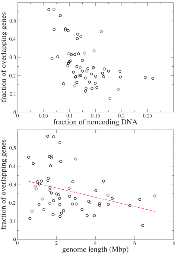
Figure 3.
Top. Scatter plot of the fraction of genes involved in (at least) an overlap versus the fraction of noncoding genome. Bottom. Scatter plot of the fraction of genes involved in (at least) an overlap versus the genome length. The dashed line is a best linear fit. Both analyses are performed by considering only overlapping genes which are both annotated in the COG database.

