
Figure 6.
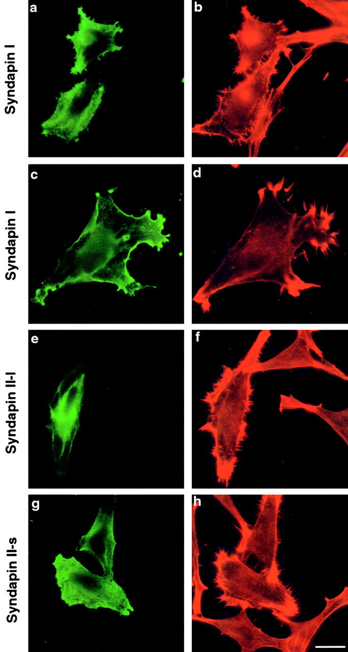
SdpI and -II induce a striking cortical actin phenotype upon overexpression. HeLa cells transiently transfected with SdpI full-length (a, b, c, and d), SdpII-l (e and f), and SdpII-s (g and h) (green fluorescence) exhibit actin microspikes (filopodia) all over the cell surfaces in addition to protrusion-like structures (lamellipodia). This overexpression phenotype was only observed in transfected cells, untransfected cells were unaffected and displayed smooth cell surfaces. Right panels show phalloidin–Texas red staining of F-actin to visualize actin rearrangements. Antitag labeling to identify syndapin-overexpressing cells is shown in the left panels. Bar: (a, b, e–h) 25 μm; (c and d) 15.75 μm.