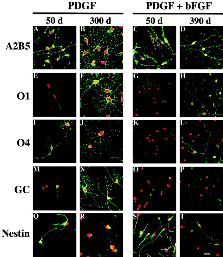
Figure 6.

Immunofluores- cence staining with A2B5, O1, O4, Ranscht anti-GC, or anti-nestin antibodies. P7 OPCs were cultured without TH in either PDGF or PDGF plus bFGF for 50 or 390 d. Antibody staining is shown in green. Nuclei were stained with propidium iodide and are shown in red. Bar, 10 μm.
