
Figure 3.
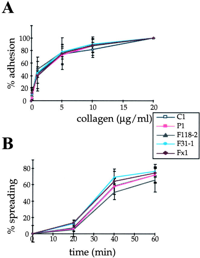
Adhesion and spreading of clones expressing wild-type and mutant forms of paxillin are similar on collagen. (A) Mock-transfected (clone C1) and cells stably transfected with wild-type (clone P1), or mutant forms of paxillin F31 (clone F31-1), F118 (clone F118-2), or F31/118 (clone Fx1) were allowed to adhere on plates coated with the indicated concentrations of collagen for 1 h at 37°C. Absorbance values corresponding to crystal violet uptake were determined for each point and expressed as percent adhesion. (B) Cell spreading was quantified by allowing cells to adhere on plates coated with 20 μg/ml collagen for the indicated times. Cell spreading was determined by calculating the percentage of spread cells at each time point. Values represent the mean of three independent experiments carried out in duplicate ± SD. Selected clones are representative of all the tested clones.