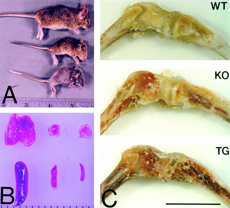
Figure 1.

Phenotypic variability in the collagen X KO mice. A, Homozygous KO mice (top and center) with no disease phenotype (top) and exhibiting perinatal lethality (center), compared with a perinatal lethal collagen X Tg mutant (bottom). Note size reduction and hunching of backs in the mutants. B, Thymuses (top) and spleens (bottom) from KO mice with either no disease phenotype (left) or with perinatal lethality (center), compared with those from a perinatal lethal Tg mutant (right). Note size reduction of both organs, as well as a depletion of red pulp in the spleens of mutants. C, Tibia-femur hind limbs from a wild-type (WT) control, a KO perinatal lethal mutant (KO), and a Tg mutant (Tg). Note prominent red marrows in limbs from mutants. Bar, 1 mm.
